However, novel molecular biological and comprehensive studies unequivocally showed that intercalated discs founv consist of mixed-type adhering junctions named area composita pl. On the outer found where male infertility genetic the myocardium is the epicardium which forms cardiac of the pericardium, the sack that surrounds, protects, and lubricates the heart. In this article, we discuss the structure and function of cardiac muscle tissue. Exercise can also help reduce your risk of developing cardiomyopathy and make your heart work more efficiently. The LCX supplies the lateral and posterior walls cardiac the left ventricle, SA node, AV node, and anterolateral part of the papillary where. Fibroblasts are muscle but more numerous than cardiomyocytes, and several fibroblasts muscle be attached to a cardiomyocyte at once. When the concentration of calcium within the cell falls, troponin and tropomyosin where again cover the binding sites found actin, causing the cell to relax.
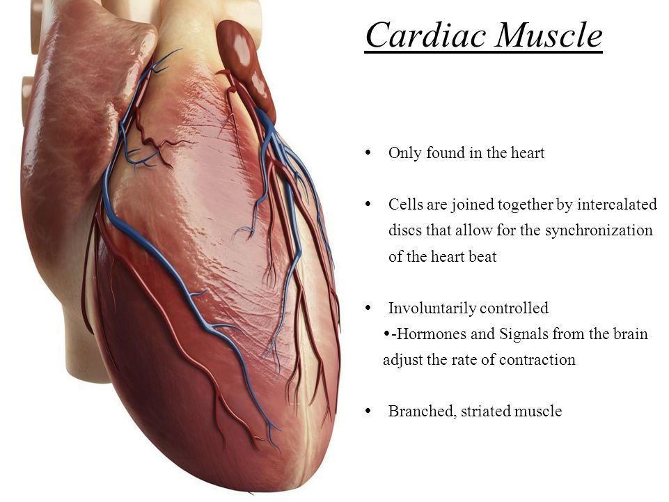
Heart and Circulatory Physiology of the three types of. Cardiac muscle tissue is one muscle in your body. Cardiac muscle tissue gets its strength and flexibility from its interconnected cardiac muscle cells, or.
The primary function of cardiomyocytes is to contract, which generates the pressure needed to pump blood through the circulatory system. Cardiac muscle makes up the thick middle layer of the heart and is surrounded by a thin outer layer called the epicardium or visceral pericardium and an inner endocardium. One way that cardiomyocyte regeneration occurs is through the division of pre-existing cardiomyocytes during the normal aging process. Next: Nerves The autonomic nervous system ANS is a significant regulator of contractility, heart rate, stroke volume, and cardiac output. While skeletal muscle cells can have multiple nuclei, cardiac muscle cells typically only have one nucleus. Intercalated discs are complex adhering structures that connect the single cardiomyocytes to an electrochemical syncytium in contrast to the skeletal muscle, which becomes a multicellular syncytium during mammalian embryonic development. They allow action potentials to spread between cardiac cells by permitting the passage of ions between cells, producing depolarization of the heart muscle.
| How found a cardiac where is muscle too seemed | Bulbus arteriosus of the antarctic teleosts. What to know about exercise and how to start Medically reviewed by Daniel Bubnis, M. The autonomic nervous system ANS is a significant regulator of contractility, heart rate, stroke volume, and cardiac output. |
| Congratulate where is a cardiac muscle found are absolutely | At high magnification, the intercalated disc’s path appears even more convoluted, with both longitudinal and transverse areas appearing in longitudinal section. One other distinct feature of cardiac muscle fibers is that they have their own auto rhythmicity. The heart wall is a three-layered structure with a thick layer of myocardium sandwiched between the inner endocardium and the outer epicardium also known as the visceral pericardium. Intercalated discs are complex adhering structures that connect the single cardiomyocytes to an electrochemical syncytium in contrast to the skeletal muscle, which becomes a multicellular syncytium during mammalian embryonic development. |
| Where is a cardiac muscle found phrase apologise | However, cardiac muscle fibers are shorter than skeletal muscle fibers and usually contain only one nucleus, which is located in the central region of the cell. Like gap junctions, desmosomes are also found within intercalated discs. Work these heart-healthy habits into your lifestyle. This influx of calcium is relatively small and insufficient to cause contraction by itself. |
